Have you had a recent fall or do you practice sports frequently and notice persistent severe pain and swelling? Did you visit a doctor and your X-rays showed nothing abnormal?
It could be a hidden fracture - let me tell you about this pain that doesn't show up on X-rays
What is a hidden fracture?
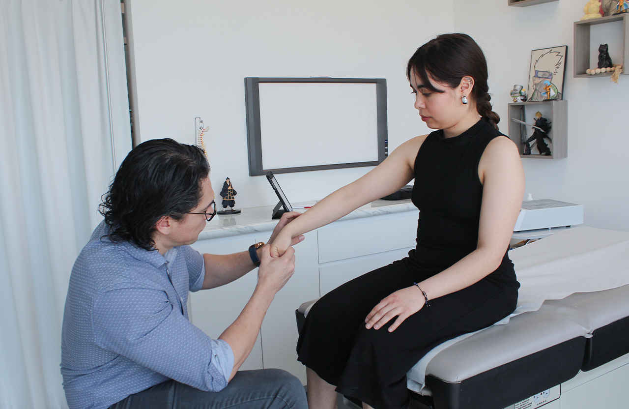
It's an injury that shows no obvious signs and therefore goes unnoticed on simple X-rays. During your physical evaluation we perform specific movements and clinical tests to locate the pain.
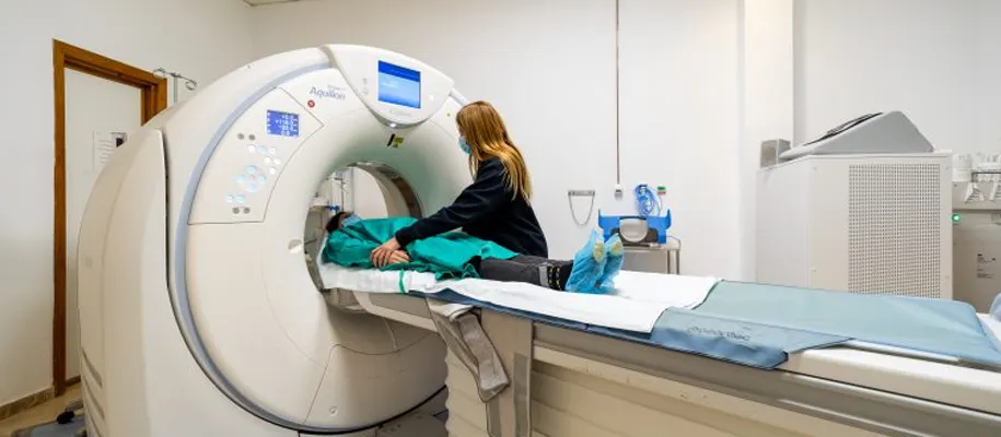
Then we request advanced imaging studies such as:
- Computed tomography (CT)
- Magnetic resonance imaging (MRI) which has proven to be a highly sensitive imaging test for diagnosing these fractures due to its high resolution.
- Bone scan which is an exam that detects bone abnormalities through injection of a radioactive tracer.
Most common areas for hidden fractures
Frequent areas include:
- Hip
- Spine
- Tibia (shinbone)
- Elbow
- Ankle
- Wrist
These last two, due to their anatomical complexity, are prone to unnoticed fractures. Women have greater predisposition due to hormonal factors like postmenopausal osteoporosis.
Hidden fractures can occur due to:
Trauma: Falls or direct impacts.
Stress fractures:
Repetitive activities (running, throwing objects, marching). Sudden increase in sports intensity that can generate micro cracks which, if not healed in time, progress to complete fractures.
Insufficiency fractures:
Weakened bones (osteoporosis, tumors, chemotherapy) that fracture with minimal effort.

Risk factors:
- Bone mass loss (osteoporosis)
- Genetic diseases (osteogenesis imperfecta)
- Rheumatoid arthritis
- Hormonal disorders (amenorrhea, menopause)
- Malnutrition or eating disorders
- Excessive alcohol or tobacco consumption
- Prolonged use of corticosteroids or anti-inflammatories
- Chronic kidney failure
Key symptoms of a hidden fracture
- Localized pain that persists even at rest
- Constant swelling
- Tenderness to touch in the affected area
- Progressive functional limitation
Treatment
Varies according to the severity and location of the fracture:
- Immobilization: Splints or cast
- Weight offloading: Crutches or canes
- Activity modification: Temporary sports rest
- Supplementation: Calcium, vitamin D and bisphosphonates in cases of osteoporosis
- Surgery: For unstable fractures or those with necrosis risk